Institute of Solid State Physics EXAFS Spectroscopy Laboratory
Institute of Solid State Physics, University of Latvia
LU CFI Webmail

ENG   LV
QR code

Google Search:


MapLarge Visitor Map

Website Statistics


Electron Microscopy

Electron microscopy is a type of microscopy that uses a beam of electrons to create an image of the specimen. There are two main types of electron microscopes: Scanning Electron Microscope (SEM) and Transmission Electron Microscope (TEM). They allow one to observe specimens in vacuum with down to atomic resolution.

In our laboratory, we use electron microscopy to study morphology and structure of nanocrystals, thin films and nanowires of different materials.
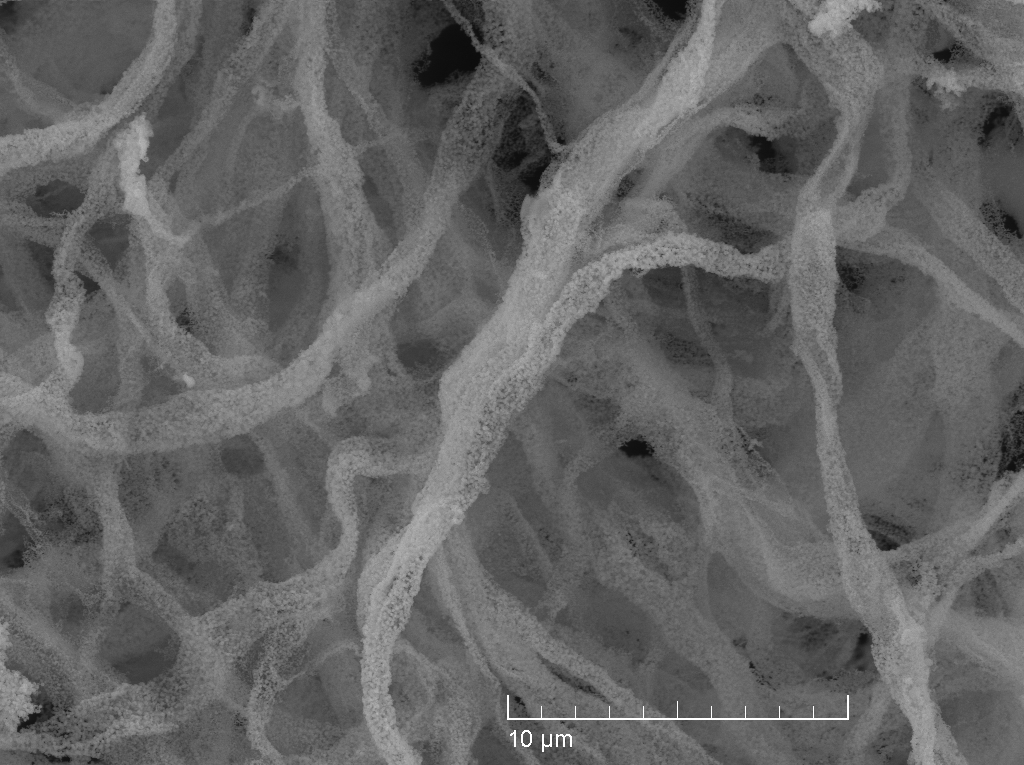
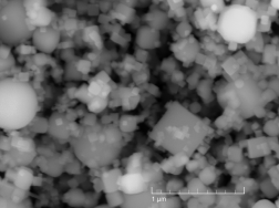
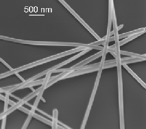
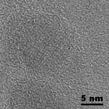


SEM image SEM image SEM image
TEM image TEM image


USEFUL LINKS

  • Electron Microscope - Wikipedia.
  • Scanning Electron Microscope - Wikipedia.
  • Transmission Electron Microscope - Wikipedia.

  • Phenom Pro - a desktop scanning electron microscope.
  • SEM-FIB Tescan Lyra XM - SEM with Schottky field emission cathode in combination with gallium Focused Ion Beam (FIB) column and with Gas Injection System (GIS).
  • TEM FEI Tecnai GF20 offers imaging and analysis solution for life sciences, materials sciences, nanotechnology, and the semiconductor and data storage industries.